無料ダウンロード venous sinuses anatomy radiology 324513-Venous sinuses anatomy radiology
Mar 13, 21 · • Dural sinuses are large, endotheliallined trabeculated venous channels encased within folds/reflections of dura that define, form their walls • Cerebral veins are thinwalled, valveless structures that cross SAS, pierce arachnoid/inner dura to enter dural venous sinus1 Department of Radiology, Queen's Medical Centre, Nottingham, UK;The more downstream venous channels (dural sinus veins) are now visualized — the superior (white) and inferior (dashed white) sagittal sinuses, the Vein of Galen (dashed yellow) and straight sinus (dashed purple), and transverse (dashed red) and sigmoid (dashed blue) sinuses, as well as the inferior petrosal sinus (dashed black)

Recanalization And Obliteration Of Falcine Sinus In Cerebral Venous Sinus Thrombosis Neurology
Venous sinuses anatomy radiology
Venous sinuses anatomy radiology-Dominant anterior cerebellar hemispheric veins (red) emptying into the superior petrosal sinus, and superior vermian vein (dark blue) opening into the Galen The superior vermian vein (dark blue) is prominent, as collector of the posterior superior surface drainage (balance, again) Inferior vermian vein is marked in orangeTHEORY OF DURAL VENOUS SINUS VIDEO https//wwwyoutubecom/watch?v=Ebw3OwEQKtcTRANS CRIBRIFORM APPROACH VIDEO https//wwwyoutubecom/watch?v=9v_C94Bwo8s&


Imaging Features Of Venous Sinus Thrombosis Part 2 Health4theworld Academy Youtube
Cavernous Sinus Thrombosis The veins of the face drain blood into the cavernous sinus via the superior ophthalmic vein As such, infections of the face (particularly those involving the "danger triangle" (orbits, nasal sinuses, and superior part of the face) can cause a cavernous sinus thrombosis Staphylococcus aureus is seen in up to 70% ofOct 22, 04 · Lateral venous phase and venous angiography showed bilateral transverse sinus stenosis (A,B), a 9 F guiding catheter was navigated with a 0018 in microwire to the proximal right jugular bulb, the microwire was passed through the stenosed transverse sinus and navigated to the opposite jugular bulb, a selfexpanded Wallsten stent of 8 × mm was deployed at the stenosisAnatomy of cerebral veins and sinuses The entire deep venous system is drained by internal cerebral and basal veins, which join to form the great vein of Galen that drains into the straight sinus Though variation in the superficial cerebral venous system is a rule, anatomic configuration of the deep venous system can be used studykorner
Dec 06, 11 · Anatomy of the dural venous sinuses;Venous sinus, in human anatomy, any of the channels of a branching complex sinus network that lies between layers of the dura mater, the outermost covering of the brain, and functions to collect oxygendepleted bloodUnlike veins, these sinuses possess no muscular coat Their lining is endothelium, a layer of cells like that which forms the surface of the innermost coat of the veinsCerebral Venous Thrombosis Radiology department of the Medical Centre Haaglanden in the Hague and the Rijnland hospital in Leiderdorp, the Netherlands Cerebral venous thrombosis is an important cause of stroke especially in children and young adults It is more common than previously thought and frequently missed on initial
Dural venous sinuses There are seven paired (transverse, cavernous, greater & lesser petrosal, sphenoparietal, sigmoid and basilar) and five unpaired (superior & inferior sagittal, straight, occipital and intercavernous) dural sinuses There are two sagittal sinuses that occupy the longitudinal cerebral fissure (midline between the cerebralOct 26, 19 · Oblique sinus ( green ) Lies behind left atrium 3 Postcaval recess ( orange ) Lies behind superior vena cava 4 Pulmonary venous recess ( red ) Left and right pulmonary venous recesses Location of pericardial sinuses from the posterior aspect of the heart Pericardial sinuses in real CT images – Transverse sinus 1In this tutorial we will review the anatomy and configuration of the dural venous sinuses We will be learning about the folllowing structures Superior sag


Cerebral Venous Sinus Thrombosis In Covid 19 Infection A Case Series And Review Of The Literature Journal Of Stroke And Cerebrovascular Diseases
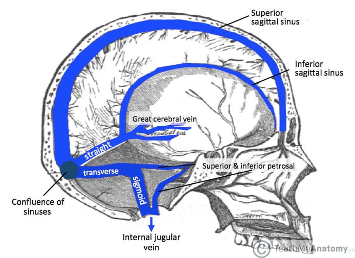


Venous Drainage Of The Cns Cerebrum Teachmeanatomy
Jun 17, 21 · Dural venous sinuses The human brain has the highest demand for oxygen of any tissue in the human body It receives a vast amount of blood from two systems the internal carotid system anteriorly, and the vertebrobasilar system posteriorly However, there is very little space in the cranial vault to accommodate large amounts of bloodFeb 25, 18 · Cerebral venous thrombosis (venous sinus thrombosis) is caused by clots in the dural venous sinuses and accounts for 05% to 1% of all strokes CVT results in an increased venous pressure that can lower cerebral perfusion pressure and induce parenchymal change due to vasogenic edema, cytotoxic edema, or intracranial hemorrhageJan 13, 19 Anatomy of cerebral veins and sinuses The entire deep venous system is drained by internal cerebral and basal veins, which join to form the great vein of Galen that drains into the straight sinus Though variation in the superficial cerebral venous system is a rule, anatomic configuration of the deep venous system can


Intracranial Venous System W Radiology


Multisection Ct Venography Of The Dural Sinuses And Cerebral Veins By Using Matched Mask Bone Elimination American Journal Of Neuroradiology
May 17, · Groen 29 The fundamental feature that distinguishes the CSVS from the systemic (caval) venous system is the lack of venous valves In 1940, Batson 2 demonstrated that the VVS was angiographically linked to the cranial venous system, and that retrograde flow from the VVS into the brain was possible because of the lack of venous valvesUniversity of Michigan Medical School Neuroanatomy Radiology University of Michigan Medical School Curricula First Year Medical Anatomy Foundations of Anatomy Anatomy Faculty and Staff M1 Foundations of Medicine II Anterior Neck and Thorax Anterior Neck and Thorax Learning Objectives Anterior Neck and Thorax LO 1Dec 01, 1994 · The normal and variant anatomy of the cerebral veins and dural venous sinuses is poorly understood by many radiologists Beginning with a discussion of cerebral venous anatomy, this review illustrates clinically pertinent anatomy of the cerebral sinovenous system Various methods of imaging cerebral veins and dural venous sinuses are described
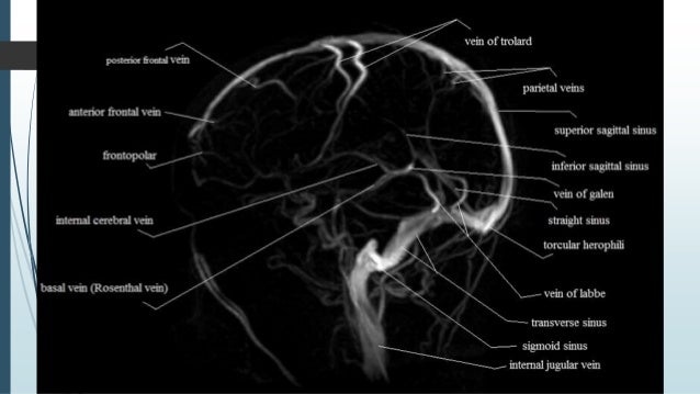


Dural Venous Sinus Thrombosis For Radiology Imaging
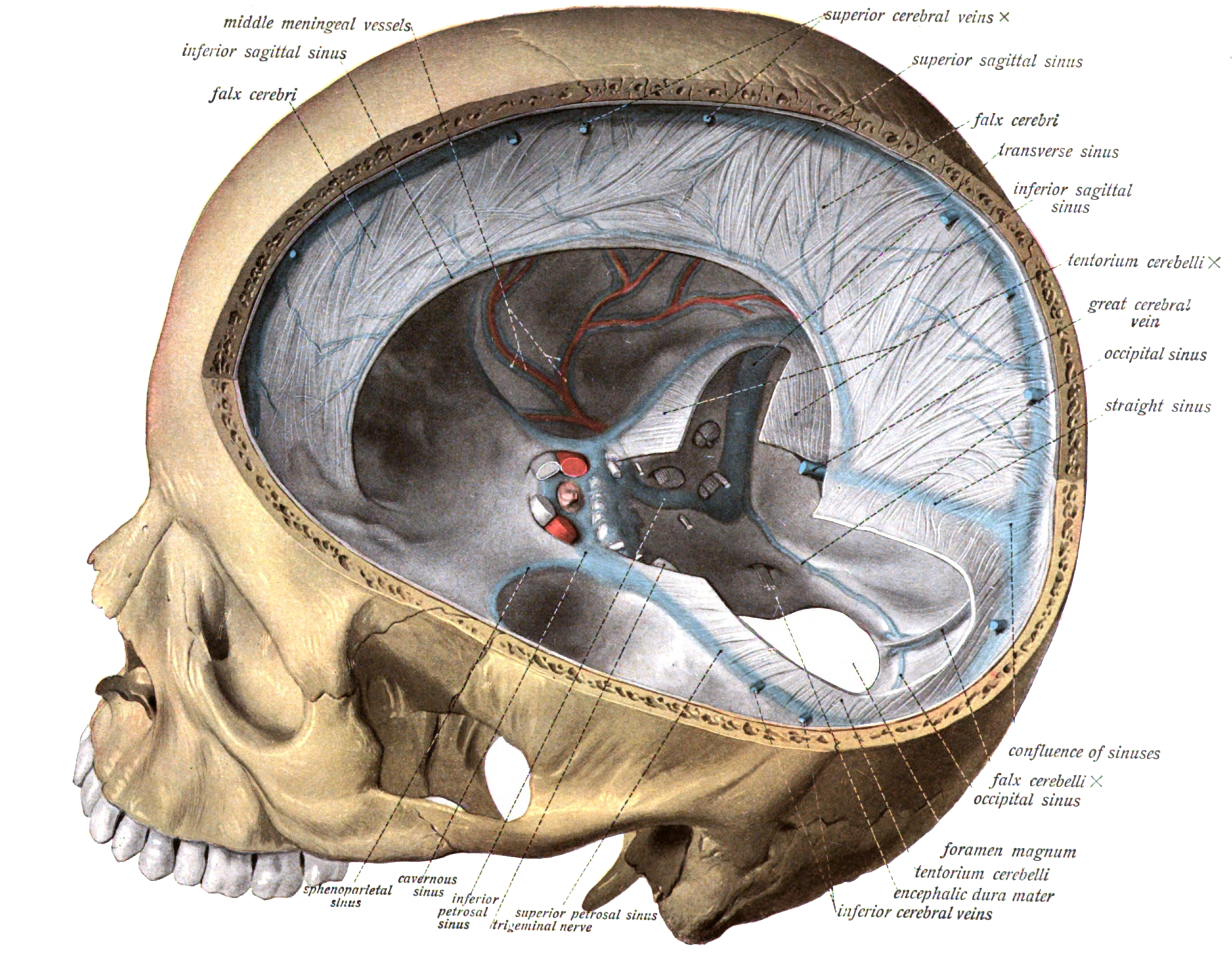


Dural Venous Sinuses Wikipedia
The intracranial or cerebral venous system is a network of nerves made up of two systems working together the superficial system and the deep system(1) The superficial cerebral system has sagittal sinuses and cortical veins The sinuses and the veins both drain deoxygenated blood from the surfaces of the brain's hemispheres(2)Dural venous sinus thrombosis is diagnosed on CT venography and MRV but CT plain brain is the initial radiological investigation Most of the time CT shows signs such as hyper density, hyperdense delta sign On this scan a subtle linear hyperdensity was seen in the region of right transverse sinus and confluence of sinusesApr 12, 13 · Variations in intracranial dural venous sinus anatomy have been widely reported in humans, but there have been no studies reporting this in dogs The purpose of this retrospective study was to describe variations in magnetic resonance (MR) venographic anatomy of the dorsal dural venous sinus system in a sample population of dogs with


Plos One Bold Granger Causality Reflects Vascular Anatomy


Cerebral Venous Sinus Sinovenous Thrombosis In Children Neurosurgery Clinics
Cerebral venous system can be divided into asuperficial and a deep system The superficial systemcomprises of sagittal sinuses and cortical veins and thesedrain superficial surfaces of both cerebral hemispheresThe deep system comprises of lateral sinus, straight sinusand sigmoid sinus along with draining deeper corticalveinsThe first of the two main divisions of this system, the intracranial veins, includes the cortical veins, the dural sinuses, the cavernous sinuses, and the ophthalmic veins The second main division, the vertebral venous system (VVS), includes the vertebral venous plexuses which course along the entire length of the spineAug 07, 19 · The venous system of the brain consists of the cerebral and cerebellar veins, which drain into the cranial venous sinuses located within the dura mater These veins are extremely thin because they lack a muscular wall, and they do not have any valves
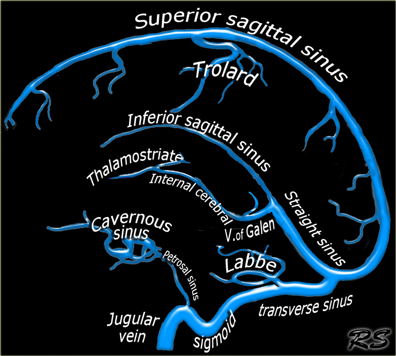


The Radiology Assistant Cerebral Venous Thrombosis
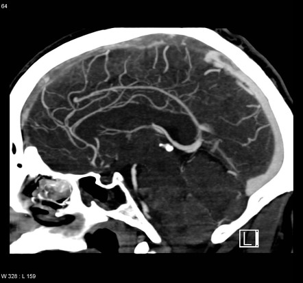


Dural Venous Sinus Thrombosis Radiology Reference Article Radiopaedia Org
Endgames Anatomy Quiz Anatomy of the dural venous sinuses BMJ 11;Anatomy, Imaging and Surgery of the Intracranial Dural Venous Sinuses, 1st Edition Editor R Shane Tubbs This firstofitskind volume focuses on the anatomy, imaging, and surgery of the dural venous sinuses and the particular relevance to neurosurgery and trauma surgery Knowledge of the fine clinical anatomy involved in neurosurgery andDiagram showing the relationship of deep cerebral veins and venous sinuses 1) Internal Cerebral Vein 2) Basal Vein of Rosenthal 3)Septal Vein 4)Thalamostriate Vein 5)


Q Tbn And9gcs Trhe8ynr 3 Tjhqiph4v9njvhhcwrmbd2dwi7rephoaofukf Usqp Cau


Paired Dural Venous Sinuses Transverse Sigmoid Superior Petrosal Inferior Petrosal Others Youtube
Craniocervical junction venous anatomy on enhanced MR images the suboccipital cavernous sinus Caruso RD(1), Rosenbaum AE, Chang JK, Joy SE Author information (1)Department of Radiology, SUNY Health Science Center, NY , USAThe superior sagittal sinus (SSS) is one of the major intradural venous sinuses, and because it is involved in various disease processes, it must be considered in intracranial neurosurgery The SSS lies in a shallow midsagittal depression at the junction of the falx cerebri, with the dura lining the inner table of the calvariaDural venous sinuses Saved by LilacLillies 168 Nervous System Anatomy Physiology Neurology Human Anatomy And Physiology Dental Anatomy Medical Science Medical Studies Brain Neurons Craniosacral Therapy


Figure 2 From Evaluation Of Dural Venous Sinuses And Confluence Of Sinuses Via Mri Venography Anatomy Anatomic Variations And The Classification Of Variations Semantic Scholar
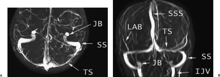


Temporal Bone Vascular Anatomy Anomalies And Disease With An Emphasis On Pulsatile Tinnitus Radiology Key
2 Department of Radiology, King's Mill Hospital, Sutton in Ashfield NG17 4JL, UK;Mar 09, 16 · Dural Venous Sinuses The dural venous sinuses lie between the periosteal and meningeal layers of the dura mater They are best thought of as collecting pools of blood, which drain the central nervous system, the face, and the scalp All the dural venous sinuses ultimately drain into the internal jugular vein Unlike most veins of the body, theJan 01, 00 · BACKGROUND AND PURPOSE MR venography is often used to examine the intracranial venous system, particularly in the evaluation of dural sinus thrombosis The purpose of this study was to evaluate the use of MR venography in the depiction of the normal intracranial venous anatomy and its variants, to assess its potential pitfalls in the diagnosis of dural venous sinus
:background_color(FFFFFF):format(jpeg)/images/article/en/dural-sinuses/MBT9Z5mR2o8TnY2hb131w_Superficial_veins_of_the_brain_lateral_view.png)


Dural Venous Sinuses Anatomy Kenhub


Normal Anatomy Of The Major Venous Sinuses On Magnetic Resonance Download Scientific Diagram
Oct 15, 17 · ARACHNOID GRANULATIONS CSFcontaining Projections That Extend From The Subarachnoid Space Into Dural Venous Sinuses Occur In All Dural Venous Sinuses(M/C Site TS And SSS And L/C SiteCS) Structure 1) A Central Core Of CSF Extends From The Subarachnoid Space Into The Granulation, 2) Which In Turn Is Covered By An Apical Cap Of Arachnoid CellsOct 07, 19 · Anatomy—The superficial venous system is made up of three groups of veins a superior group, comprising the superior cerebral veins and the vein of Trolard, which connects the superficial middle cerebral vein with the superior sagittal sinus;We have termed this the venous distension sign


Cerebral Venous Sinus Thrombosis On Mri A Case Series Analysis


Radiologic Clues To Cerebral Venous Thrombosis Radiographics
Venous sinus anatomy appeared unusual, and thus magnetic resonance venography was performed, which identified the OS as the main drainage pathway for the entire brain, providing the sole drainage between the superior sagittal sinus and the jugular veins through the marginal sinus Both the transverse and sigmoid sinuses were hypoplastic, andThe dural venous sinuses (also called dural sinuses, cerebral sinuses, or cranial sinuses) are venous channels found between the endosteal and meningeal layers of dura mater in the brainBACKGROUND AND PURPOSE Patients with intracranial hypotension (IH) demonstrate intracranial venous enlargement with a characteristic change in contour of the transverse sinus seen on routine T1weighted sagittal imaging In IH, the inferior margin of the midportion of the dominant transverse sinus acquires a distended convex appearance;


Mrv Major Venous Sinuses Are Labeled With Arrows Download Scientific Diagram
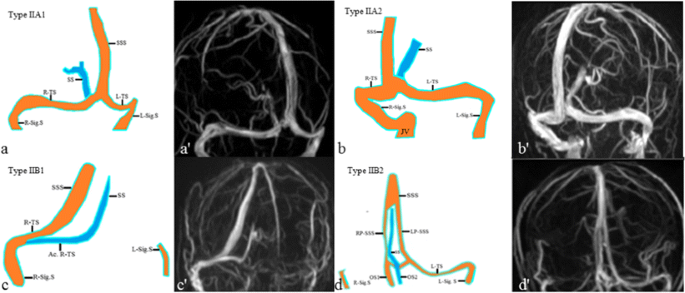


Evaluation Of Dural Venous Sinuses And Confluence Of Sinuses Via Mri Venography Anatomy Anatomic Variations And The Classification Of Variations Springerlink
Dural venous sinus thrombosis (plural thromboses) is a subset of cerebral venous thrombosis, often coexisting with cortical or deep vein thrombosis, and presenting in similar fashions, depending mainly on which sinus is involved As such, please refer to the cerebral venous thrombosis article for a general discussionAnd an inferior group, comprising the inferior cerebral veins and the vein of Labbé (which connects the middle cerebral vein with the transverse sinus)Cerebral venous sinus thrombosis (CVST) occurs when a blood clot forms in the brain's venous sinuses This prevents blood from draining out of the brain As a result, blood cells may break and leak blood into the brain tissues, forming a hemorrhage This chain of events is part of a stroke that can occur in adults and children


Http Pdf Posterng Netkey At Download Index Php Module Get Pdf By Id Poster Id


Car Ca Uploads Education lifelong learning Meetings Asm15 Speakers Pres Ee030 Imaging Approach To Cerebral Venous Thrombosis Dmytriw Pdf
Generally, venous blood drains to the nearest venous sinus, except in the case of that draining from the deepest structures, which drain to deep veins These drain, in turn, to the venous sinuses The superficial cerebral veins can be subdivided into three groups These are interlinked with anastomotic veins of Trolard and LabbeMay 02, 17 · • Drainage of the cavernous sinus is via – superior petrosal sinus to the transverse sinus – inferior petrosal sinus directly to the jugular bulb – venous plexus on the internal carotid artery to the basilar venous plexuses – emissary viens passing through the • sphenoidal foramen • foramen ovale • foramen lacerum • Additionally the cavernous sinuses connect to each other via the intercavernous sinusesA medial group, comprising the superficial middle cerebral vein;
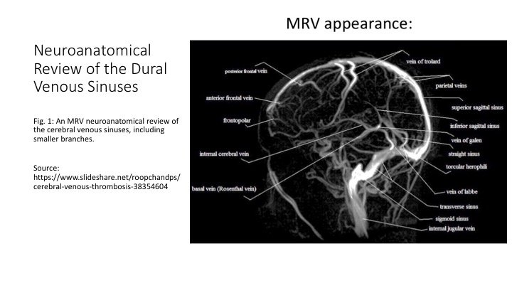


Epos C 1049
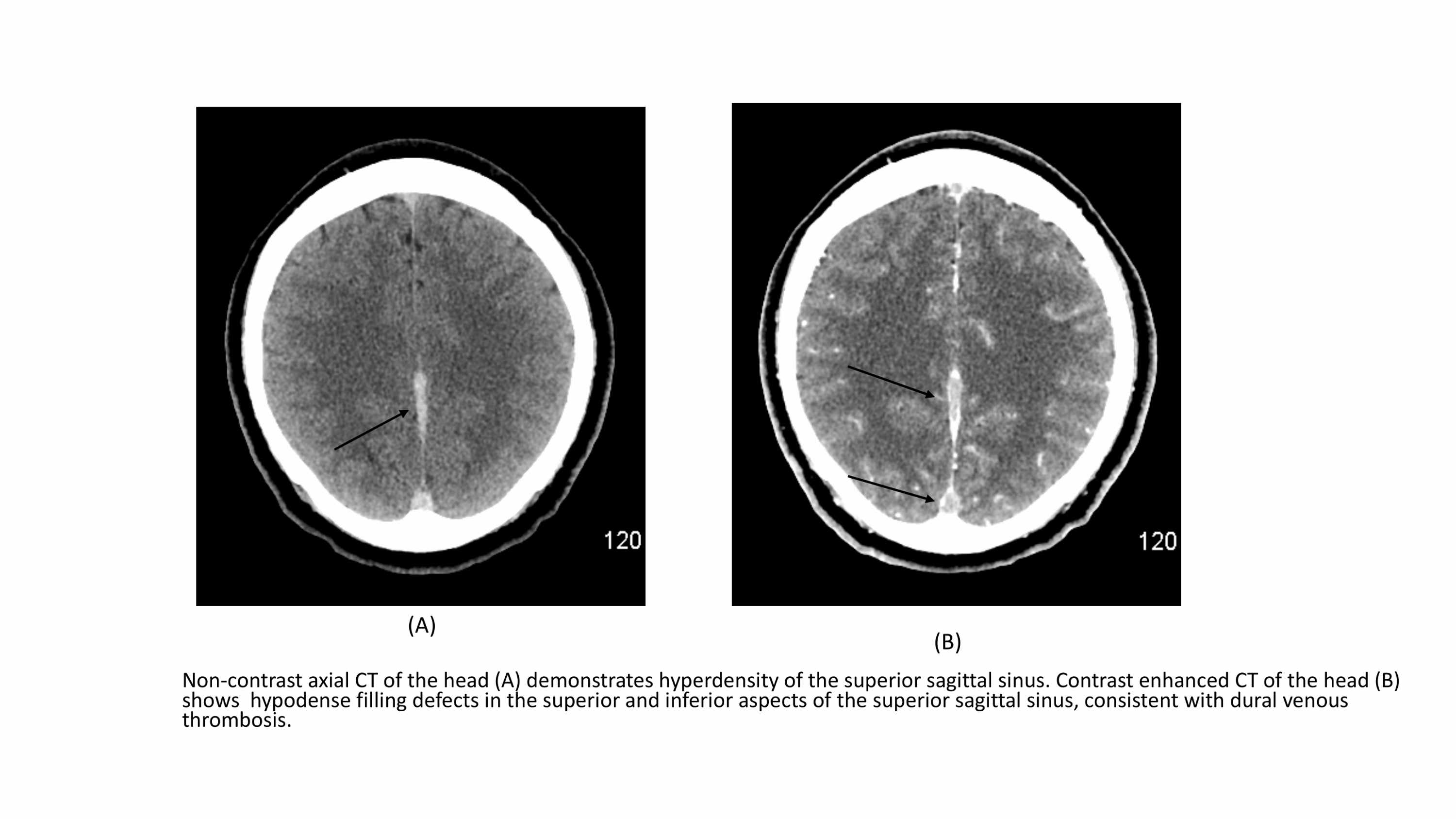


Cureus Cerebral Venous Sinus Thrombosis In Covid 19 An Unusual Presentation
Knowledge of dural venous sinus anatomy and pathomechanisms of injury will aid the radiologist in prompt recognition of venous injury, regardless of whether imaging was tailored for the evaluation of the dural venous sinuses Learning Objective Briefly review dural venous sinus anatomy and describe the imaging appearance and pathomechanisms of
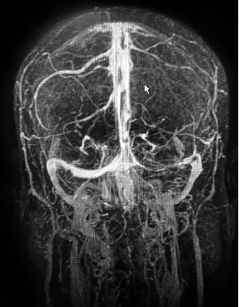


How To Spot And Treat Cerebral Venous Sinus Thrombosis Acep Now


Radiological Imaging Of Cerebral Venous Thrombosis Ppt Video Online Download


Diagnostic Performance Of Routine Brain Mri Sequences For Dural Venous Sinus Thrombosis American Journal Of Neuroradiology


Slow Flow V Thrombus Questions And Answers In Mri
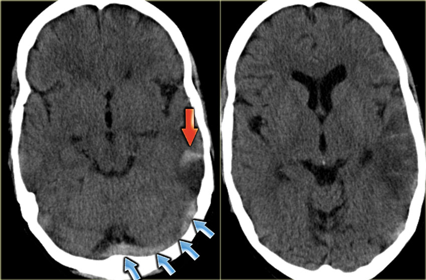


The Radiology Assistant Cerebral Venous Thrombosis


Intracranial Venous System W Radiology
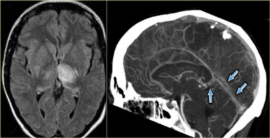


The Radiology Assistant Cerebral Venous Thrombosis


Slow Flow V Thrombus Questions And Answers In Mri


Intracranial Venous System W Radiology
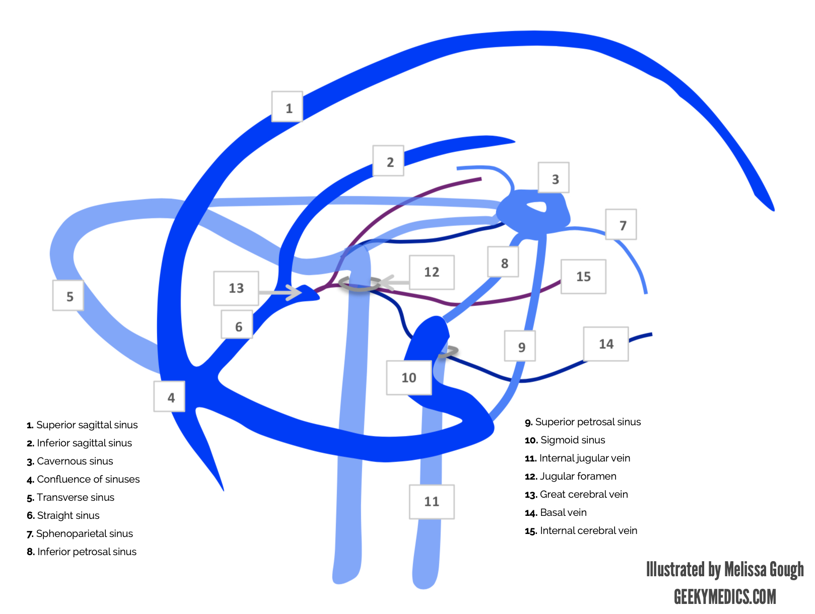


Venous Drainage Of The Brain Anatomy Geeky Medics


Dural Venous Sinuses 3d Anatomy Tutorial Youtube
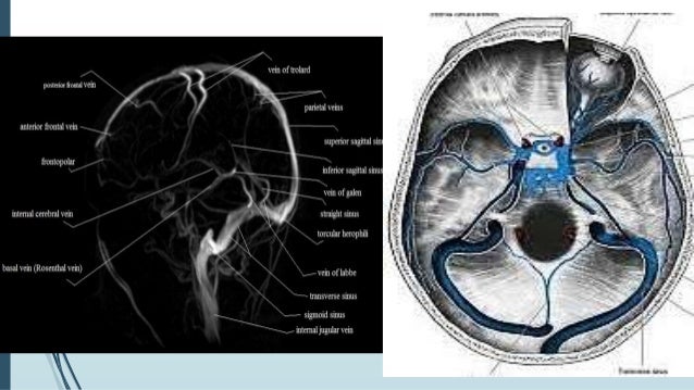


Dural Venous Sinus Thrombosis For Radiology Imaging


Radiologic Clues To Cerebral Venous Thrombosis Radiographics


Dural Venous Sinus Thrombosis Radiology Reference Article Radiopaedia Org


Dural Venous Sinuses Wikipedia


Cerebral Venous Thrombosis A Practical Guide Practical Neurology


Brain Veins And Venous Sinuses Mri Angiogram Stock Image C048 9053 Science Photo Library
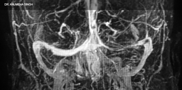


How To Spot And Treat Cerebral Venous Sinus Thrombosis Acep Now


Recanalization And Obliteration Of Falcine Sinus In Cerebral Venous Sinus Thrombosis Neurology
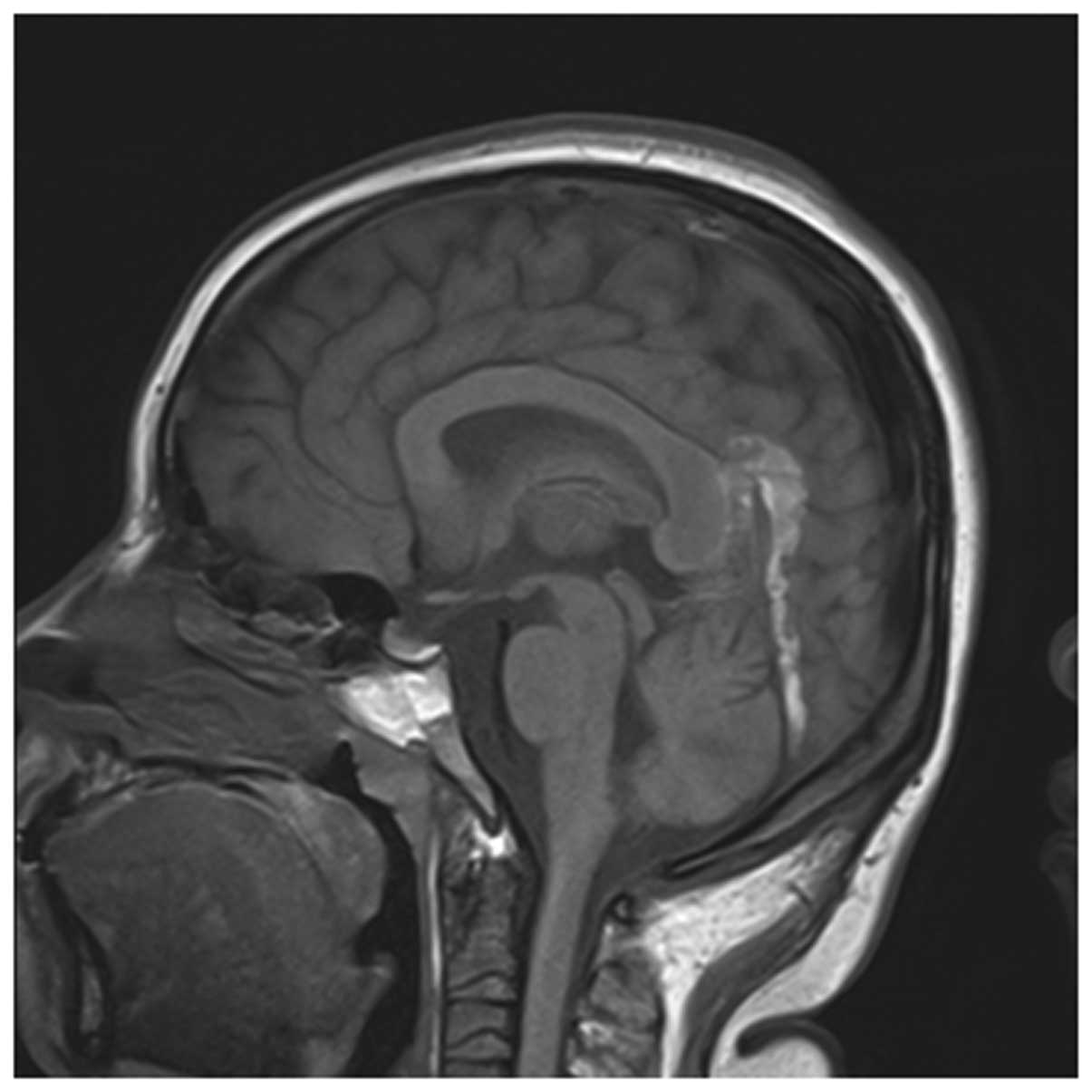


Early Imaging Characteristics Of 62 Cases Of Cerebral Venous Sinus Thrombosis


Figure 2 From Diagnostic Performance Of Mri Sequences For Evaluation Of Dural Venous Sinus Thrombosis Semantic Scholar


Normal Structures In The Intracranial Dural Sinuses Delineation With 3d Contrast Enhanced Magnetization Prepared Rapid Acquisition Gradient Echo Imaging Sequence American Journal Of Neuroradiology
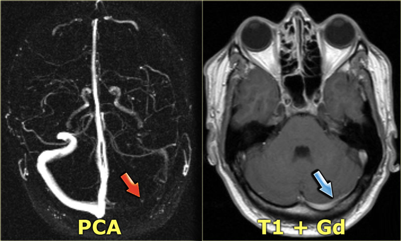


The Radiology Assistant Cerebral Venous Thrombosis


Imaging Features Of Venous Sinus Thrombosis Part 2 Health4theworld Academy Youtube
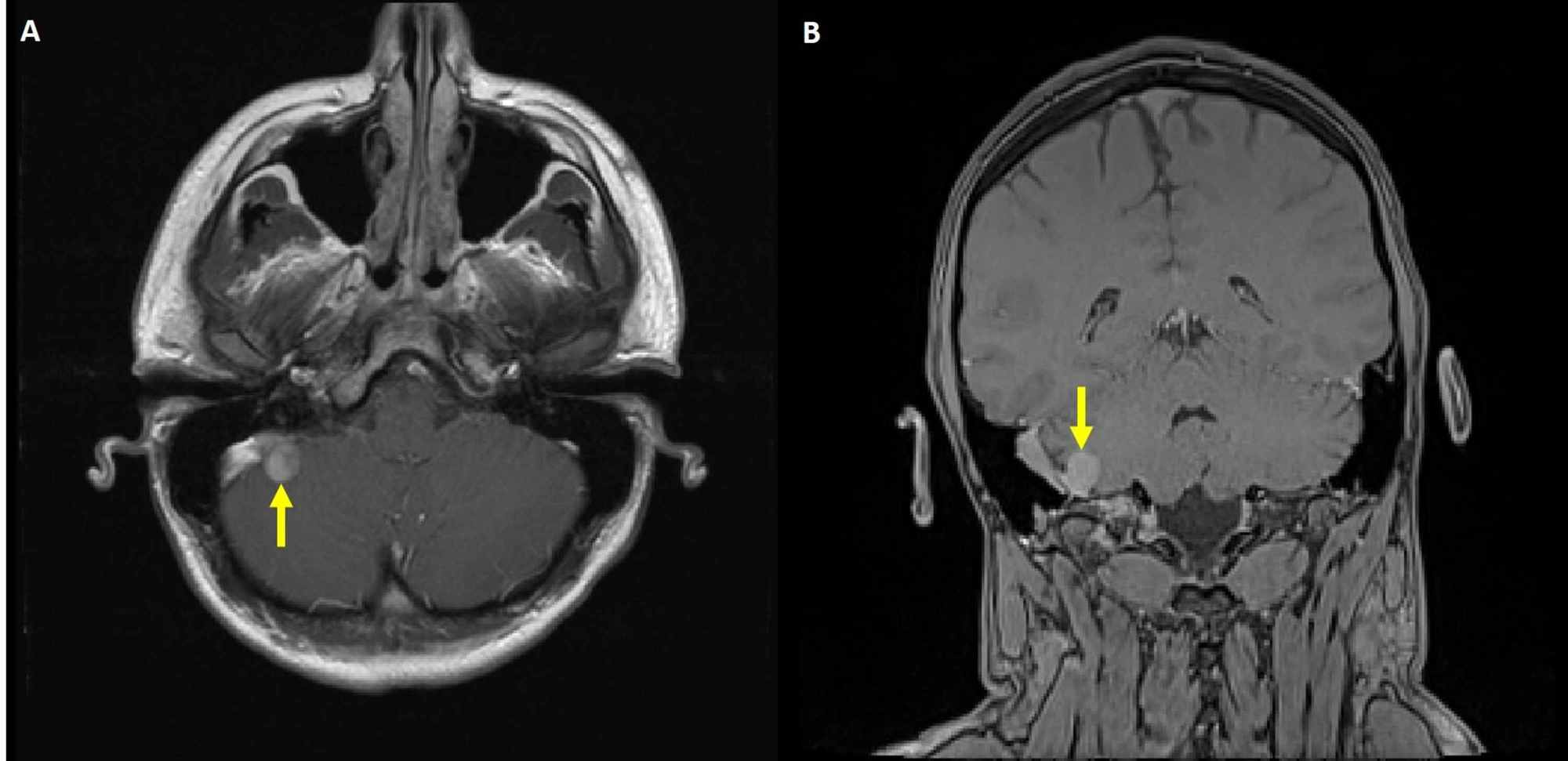


Cureus Venous Manometry As An Adjunct For Diagnosis And Multimodal Management Of Intracranial Hypertension Due To Meningioma Compressing Sigmoid Sinus
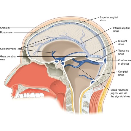


Dural Venous Sinuses Radiology Reference Article Radiopaedia Org


Dural Venous Sinus Thrombosis For Radiology Imaging
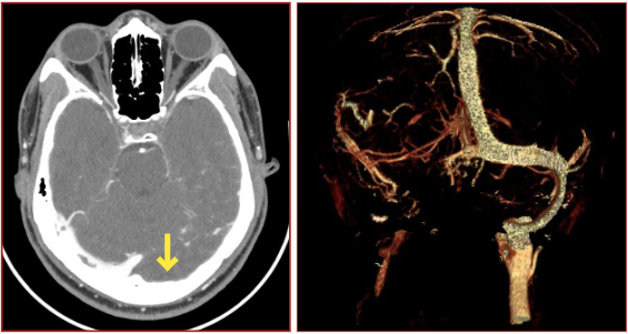


Annals Of B Pod Dural Venous Sinus Thrombosis Taming The Sru
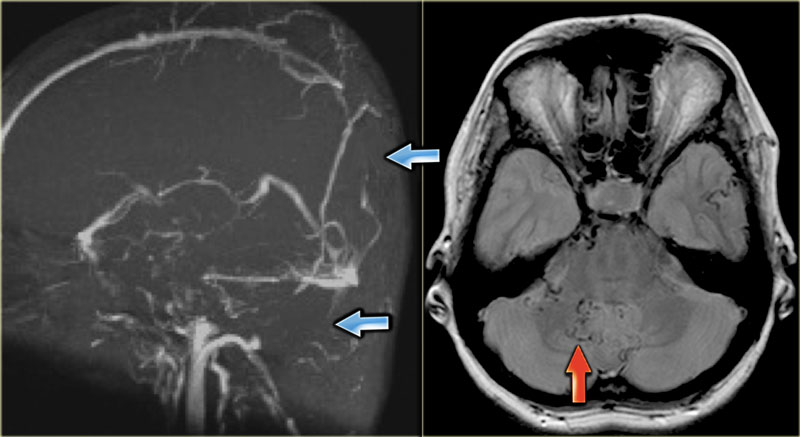


The Radiology Assistant Cerebral Venous Thrombosis
/2020/images/figure1.1586244378.jpg)


Papilledema And Diplopia Due To Meningioma Inside The Superior Sagittal Sinus A Case Report And Review Of The Literature International Journal Of Case Reports And Images Ijcri


Management Of Meningiomas Involving The Transverse Or Sigmoid Sinus In Neurosurgical Focus Volume 35 Issue 6 13
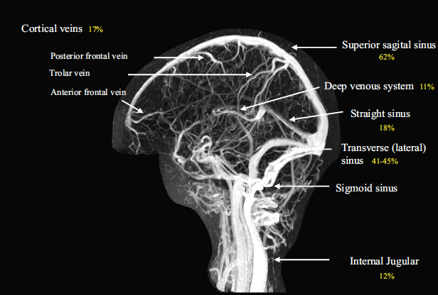


Cerebral Venous Thrombosis Core Em


Q Tbn And9gcrf1r8ogr0cp0oo znfe9nz8qwplldxkggncyb03hfu Nwpbj Usqp Cau


1


1


Anatomy Of Cerebral Veins And Dural Sinuses Sciencedirect


Venous Sinuses Neuroangio Org


Anatomy Of The Dural Venous Sinuses The Bmj
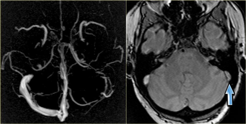


The Radiology Assistant Cerebral Venous Thrombosis


Radiologic Clues To Cerebral Venous Thrombosis Radiographics
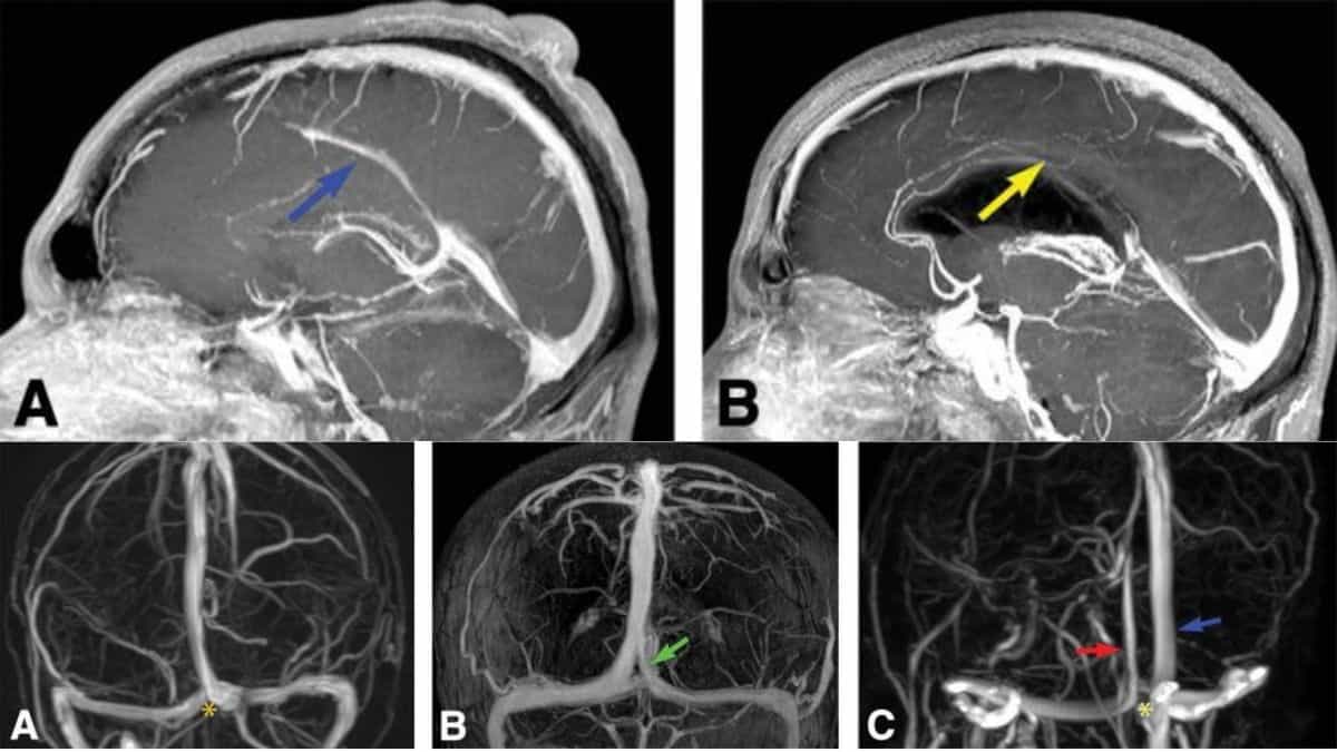


Normal As Well As Variant Anatomy Of The Dural Venous Sinuses Woms


Figure 4 From Mri Diagnosis Of Dural Sinus Cortical Venous Thrombosis Immediate Post Contrast 3d Gre T1 Weighted Imaging Versus Unenhanced Mr Venography And Conventional Mr Sequences Semantic Scholar
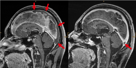


Cerebral Vein And Sinus Thrombosis
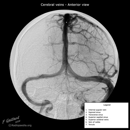


Transverse Sinus Radiology Reference Article Radiopaedia Org


Early Detection And Quantification Of Cerebral Venous Thrombosis By Magnetic Resonance Black Blood Thrombus Imaging Stroke


Pulsatile Tinnitus Venous Sinus Stenosis And Stenting Neuroangio Org


Cerebral Venous Thrombosis Something About Radiology Just For Sharing


Dural Venous Sinuses Radiology Reference Article Radiopaedia Org


Imaging Of Cerebral Venous Thrombosis Sciencedirect
%20magnetic%20resonance%20venogram%20anatomy%20image%20coronal.jpg)


Veins Of Brain Mrv Brain Veins Anatomy


View Of Bilateral Transverse Sinus Hypoplasia A Rare Case Report Journal Of Experimental And Clinical Neurosciences


Venous Sinuses Neuroangio Org


Venous Sinuses Neuroangio Org


Intracranial Venous System W Radiology


Dural Venous Sinuses Anatomy Kenhub


Teaching Neuroimages Congenital Variant Misdiagnosed As Cerebral Venous Sinus Thrombosis Neurology
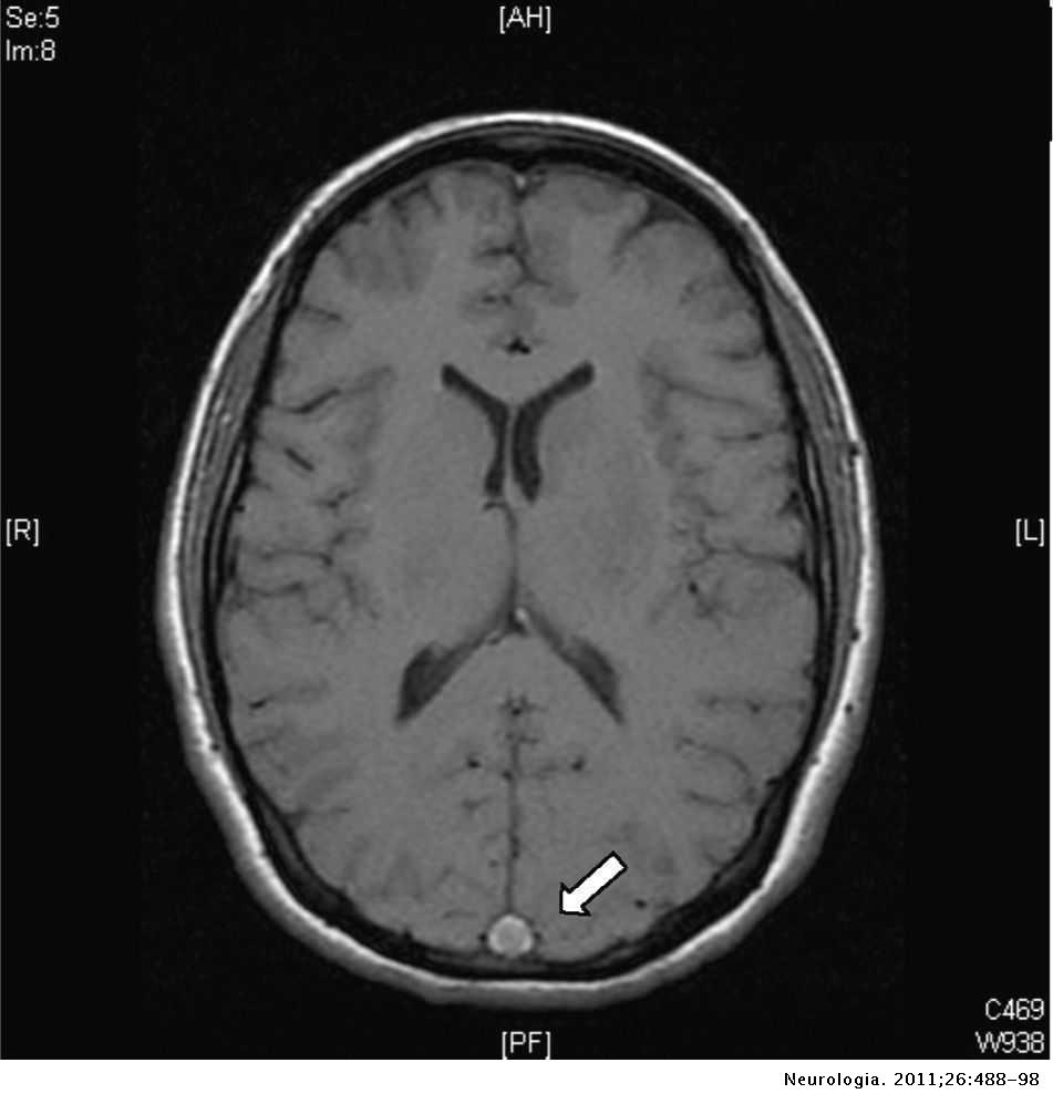


Cerebral Venous Thrombosis A Diagnostic And Treatment Update Neurologia English Edition
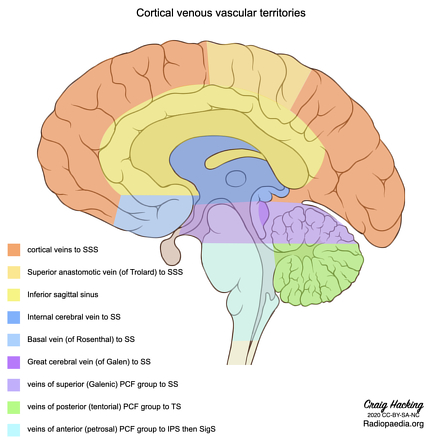


Dural Venous Sinus Thrombosis Radiology Reference Article Radiopaedia Org


Cerebral Venous Thrombosis Something About Radiology Just For Sharing


Venous Sinus Thrombosis Radiology At St Vincent S University Hospital
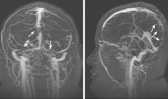


Cerebral Venous Thrombosis Radiology Key


A Review Of Extraaxial Developmental Venous Anomalies Of The Brain Involving Dural Venous Flow Or Sinuses Persistent Embryonic Sinuses Sinus Pericranii Venous Varices Or Aneurysmal Malformations And Enlarged Emissary Veins In Neurosurgical


Pictorial Review Of Intracranial Mrv Techniques Pitfalls And
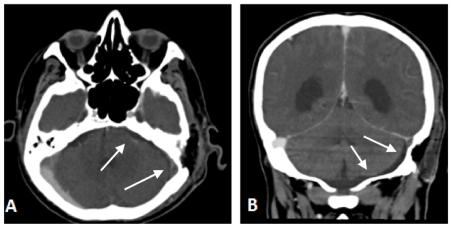


Septic Thrombophlebitis Of The Sigmoid Sinus After Vestibular Schwannoma Resection A Case Report And Literature Review


Dural Venous Sinus Thrombosis For Radiology Imaging


Venous Sinuses Neuroangio Org
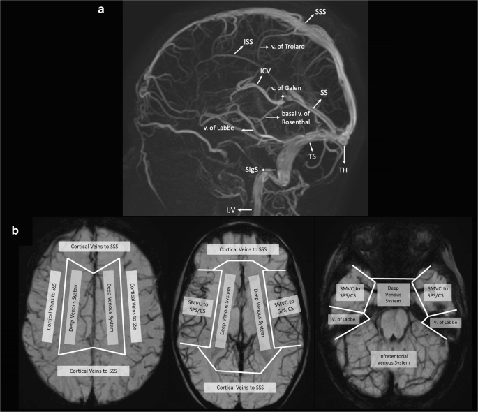


Venous Pathologies In Paediatric Neuroradiology From Foetal To Adolescent Life Springerlink


Figure 1 From Cerebral Sinovenous Thrombosis Csvt In Children What The Pediatric Radiologists Need To Know Semantic Scholar
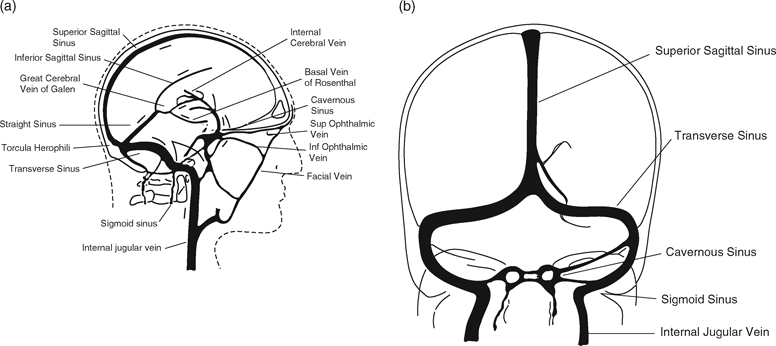


Cerebral Venous Thrombosis And Intracerebral Hemorrhage Chapter 7 Intracerebral Hemorrhage


Radiological Imaging Of Cerebral Venous Thrombosis Ppt Video Online Download
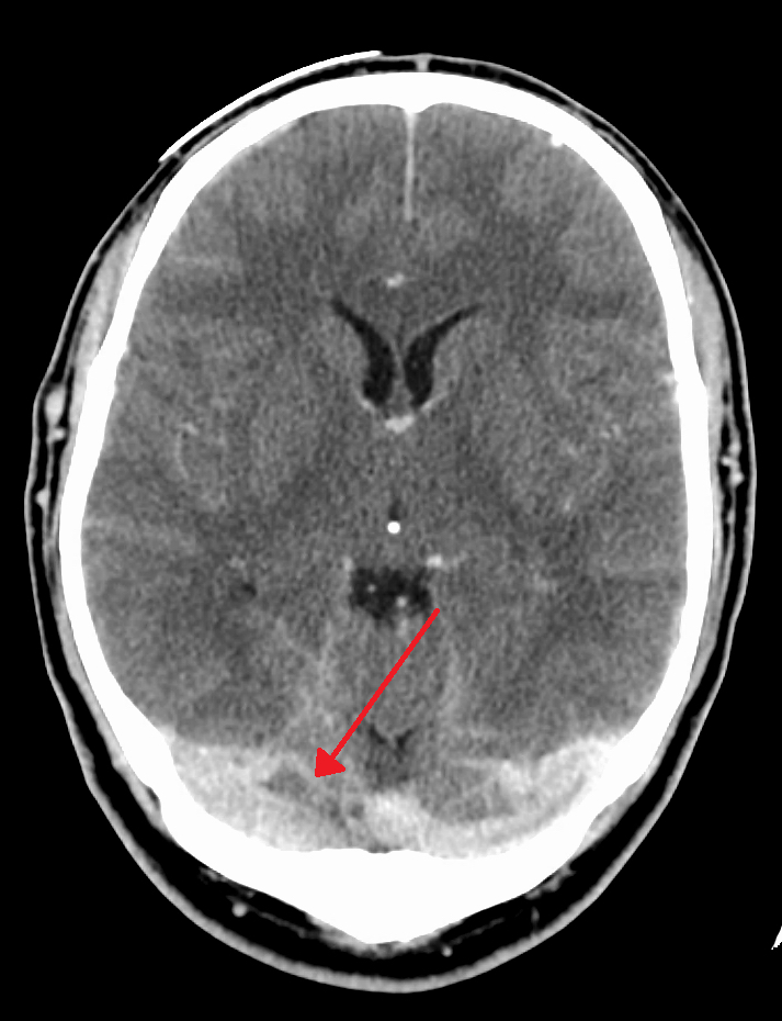


Venous Drainage Of The Brain Anatomy Geeky Medics


Imaging Of Cerebral Venous Thrombosis Clinical Radiology


Cerebral Mr Venography Normal Anatomy And Potential Diagnostic Pitfalls American Journal Of Neuroradiology
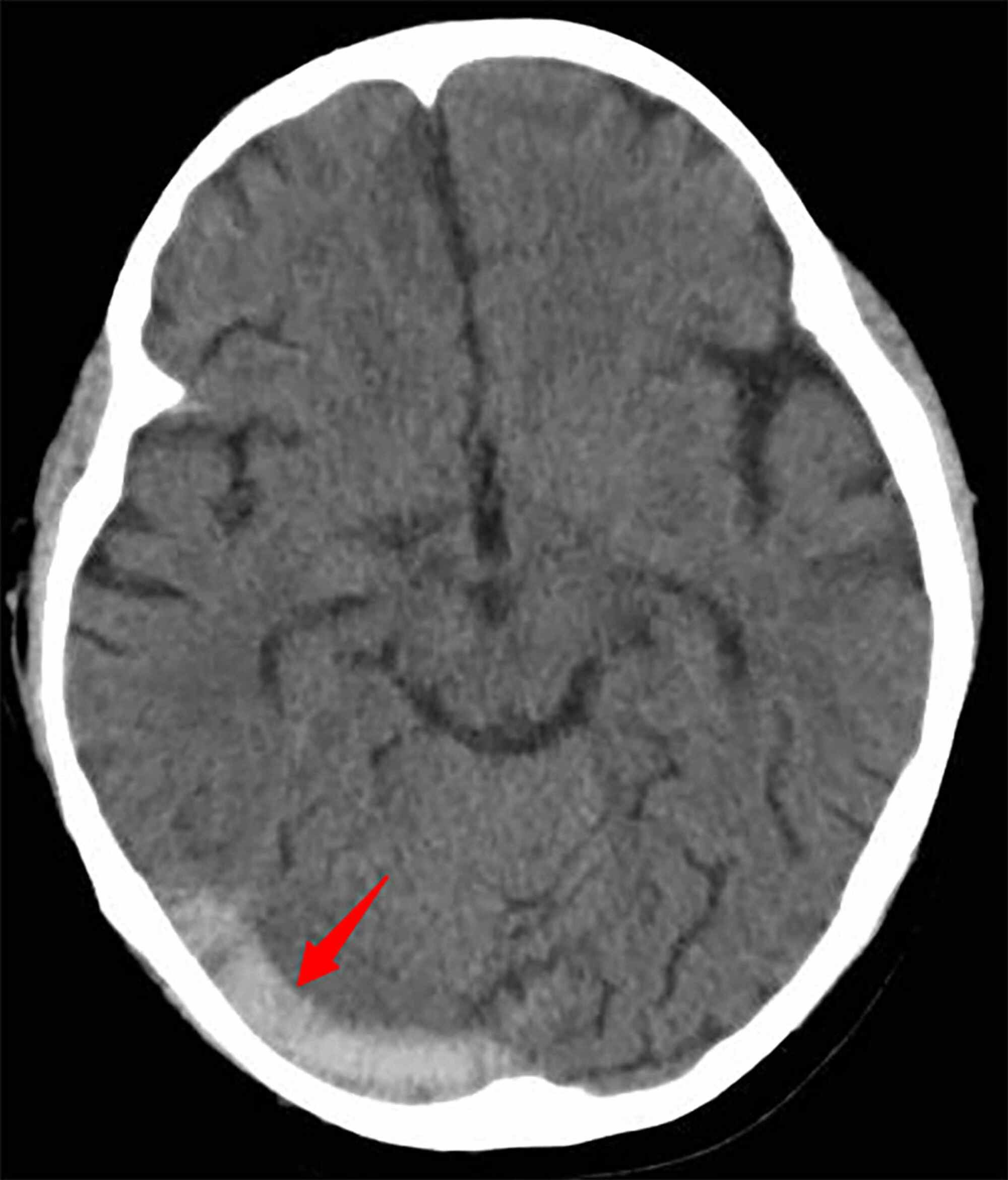


Cureus Cerebral Venous Sinus Thrombosis In A Child With Idiopathic Nephrotic Syndrome A Case Report And Review Of The Literature


Cerebral Venous Thrombosis Emcrit Project


Current Endovascular Strategies For Cerebral Venous Thrombosis Report Of The Snis Standards And Guidelines Committee Journal Of Neurointerventional Surgery
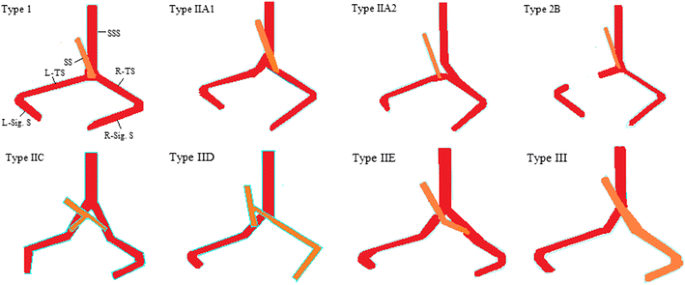


Evaluation Of Dural Venous Sinuses And Confluence Of Sinuses Via Mri Venography Anatomy Anatomic Variations And The Classification Of Variations Springerlink


Occipital Sinus It Is The Smallest Of The Dural Venous Sinuses Andlies On The Inner Surface Of The Occipital Bone F Sinusitis Radiology Occipital
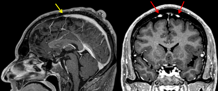


Epos C 0556


Pin On Snc


コメント
コメントを投稿